• A new study led by the Centro Nacional de Análisis Genómico (CNAG) reveals how cerebrospinal fluid contains key information on the progression of central nervous system neoplasms and their resistance to treatments.
• Published in Cell Reports Medicine, the research combines, for the first time, cerebrospinal fluid liquid biopsy with a suite of next-generation sequencing techniques.
• The findings open new avenues for developing personalised therapies for patients with leptomeningeal diseases, characterised by the presence of cancer cells in the cerebrospinal fluid.
March 09, 2026. Every day, our brain is bathed and cleansed with around half a litre of a colourless fluid: the cerebrospinal fluid. This “brain plumbing system” penetrates the deepest layers of the nervous system and carries highly valuable information about what is happening at its epicentre, including immune cell activity. Until now, cerebrospinal fluid has primarily been used to diagnose various neurological diseases via lumbar puncture, helping thousands of patients understand their condition.

Now, the Centro Nacional de Análisis Genómico (CNAG), in collaboration with
Goethe University Frankfurt (Germany), has taken a step further by
combining these samples with a suite of next-generation sequencing techniques to decode the cerebrospinal fluid and obtain key insights into the evolution of brain tumours and their treatment resistance. The study, published in
Cell Reports Medicine, analyses immune system activity in the cerebrospinal fluid, producing a high-resolution image of immune cell types and their interactions in patients with central nervous system neoplasms.
“Studying brain tumours has always been a major challenge, especially due to the difficulty of accessing them,” explains Dr Holger Heyn, lead author of the study, ICREA Professor and Head of the Single-Cell Genomics Group at CNAG. “In this study, thanks to the cerebrospinal fluid, we’ve dived into the brain’s own plumbling and discovered a true hidden gem: direct information on tumour evolution and its dialogue with the immune system. This finding makes cerebrospinal fluid a powerful prognostic tool, allowing us to uncover previously unknown biomarkers that could form the basis for designing more effective therapies.”
Decoding the tumour microenvironment of the brain
Although the brain is an immune-privileged organ, our immune cells still migrate to it to fight tumours. As with any neurological disease, the presence of these cells in cerebrospinal fluid increases significantly in patients as the disease progresses, aiming to reach the epicentre and combat it.
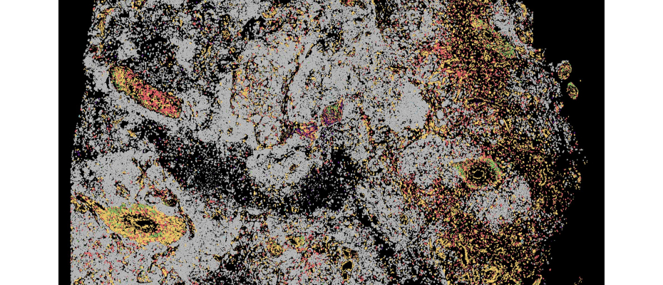
The researchers analysed over 70,000 cells from patients with cerebrospinal fluid lymphomas, glioblastomas, or brain metastases from melanoma, breast, and lung cancer, comparing them with cells from patients with neuroinflammatory disorders. CNAG’s team used pioneering sequencing techniques that allowed the RNA of each cerebrospinal fluid cell to be studied individually, revealing how genes are activated and how cells respond to the tumour, including T-cell receptors, which act as “sensors” to detect and attack cancer, with their expansion reflecting the strength of the immune response. The analysis was complemented with tumour DNA to identify potential vulnerabilities and spatial transcriptomics to produce a detailed map of cells in their environment.
“Thanks to this innovative approach, we’ve learned that each tumour type creates its own microenvironment in the cerebrospinal fluid, reflecting what may be occurring at the disease’s epicentre,” explains Dr Juan Nieto, lead author and CNAG immunologist. “This helps us better understand tumour behaviour and immune response, providing very useful information for developing new tools to monitor tumour dynamics.”
A step forward for personalised therapies
Given the aggressiveness of central nervous system neoplasms and the limited effectiveness of current therapies, the scientific community is eager to better understand the biology of these tumours and the body’s defence mechanisms. In this urgent race for answers, this study represents a major step forward, highlighting new potential biomarkers for designing more effective personalised therapies.
“Through these new analyses, we’ve been able to identify T cells with specific receptors capable of recognising tumour cells in cerebrospinal fluid, whose possible entry route is through the bloodstream” explains Paula Nieto, first author of the study and CNAG researcher. “This could allow the design of personalised therapies, such as T cells engineered with specific receptors or targeted vaccines, harnessing the body’s own defence to fight cancer. While much work remains, these strategies could mark an important advance in brain tumour treatment, contributing to more precise therapies.”
These immunotherapies exploit the patient’s own T cells, training or modifying them to recognise and attack the tumour more effectively. Among the most advanced options are TCR-T cells (T cells modified with specific receptors), CAR-T cells designed to detect tumour antigens, and receptor-based vaccines, which aim to stimulate the immune system to act directly against cancer.
Currently, the study is in its second phase, applying this innovative approach to an expanded cohort of patients to analyse immune responses before and after treatment, with the ultimate goal of making this new methodology a universal analytical tool, scalable for use in hospitals and medical centres worldwide.
GRANTS
The human spatial transcriptomics brain data were generated using the Xenium Analyzer, a state-of-the-art instrument available at CNAG. The acquisition of this technology was supported by grant ICTS2022-007832, funded by MICIU/AEI/10.13039/501100011033 and the European Union NextGenerationEU/PRTR.
REFERENCE ARTICLE
Nieto, Paula, et al. ‘Integrative CSF Profiling Identifies Disease-Specific Immune Responses in Leptomeningeal Disease’. Cell Reports Medicine, Mar. 2026, p. 102651. DOI.org (Crossref),
https://doi.org/10.1016/j.xcrm.2026.102651.