Centre Nacional d'Anàlisi Genòmica
Centro Nacional de Análisis Genómico
Centro Nacional de Análisis Genómico
- Researchers from the Centro Nacional de Análisis Genómico (CNAG, Spain), together with the St. Jude Children’s Research Hospital (USA) and the University of Adelaide (Australia), have developed a revolutionary technique called STAMP, which analyses millions of individual cells at once by leveraging state-of-the-art spatial omics imaging platforms, thus bypassing the need for sequencing.
- This novel method, published in Cell, significantly reduces analysis time and cost, while increasing the number of cells studied simultaneously from thousands to millions, offering a new and affordable approach to single-cell analysis for research laboratories and pharmaceutical organisations.
- By applying STAMP, researchers can capture snapshots of individual cells in suspension, providing morphological information together with high-resolution profiles of their transcriptome and proteome that reveal gene expression and protein function—key to improving diagnosis, guiding drug development, and advancing precision medicine.
Until now, decoding the complexity of cells within the blood, tissues and organs meant reading millions of letters — A, G, C, and T — by sequencing and assembling them into an instruction book that reveals how a cell functions, whether healthy or diseased. But a revolutionary technique transformed single-cell analysis forever. For the first time, scientists can capture high-resolution snapshots of cells directly from liquid samples—without the need for sequencing. This novel method, called STAMP, takes its name from the process of ‘stamping’ liquid samples as if they were tissue. Published today in the prestigious journal Cell, STAMP was developed by an international team from the Centro Nacional de Análisis Genómico (CNAG, Spain), St. Jude Children’s Research Hospital (USA), and the University of Adelaide (Australia).
The full name of this groundbreaking technique—Single-Cell Transcriptomics Analysis and Multimodal Profiling through Imaging (STAMP)— reveals its key innovations. First, the method enables the study of individual cells from a wide range of sources, including blood samples (such as liquid biopsies), cancer cells, embryonic stem cells, either in the form of whole cells or isolated nuclei. Second, STAMP allows researchers to analyse the transcriptome (the RNA molecules that show which genes are active) and the proteome (the proteins that define how a cell functions), either independently or at the same time. Finally, the technique captures this molecular information by imaging the samples—taking snapshots using spatial genomics technologies—instead of sequencing them, additionally giving information on the shape and morphology of the cells.
According to Dr. Holger Heyn, Single-Cell Genomics Group Leader at CNAG and one of the authors of this method: “STAMP can be a game-changer for understanding complex diseases like cancer, neurodegenerative disorders, and autoimmune conditions. By revealing critical changes in cell morphology, RNA and protein profiles of millions of cells or hundreds of samples, STAMP uncovers hidden clues about disease biology and treatment response that were previously impossible to detect. With this technology we open the door to revolutionary advances in precision medicine, enabling the development of highly targeted diagnostics and therapies that could transform patient outcomes”.
One of the key contributions of STAMP lies in making single-cell analysis faster and more accessible for research laboratories and pharmaceutical organisations. This method provides important molecular information that was difficult to obtain with traditional approaches, such as identifying ultra-rare populations like circulating tumor cells (CTCs), which are crucial for understanding cancer metastasis. STAMP also enables large-scale studies involving gene editing or drug testing in lab models, as well as detailed immune profiling of cells — all essential information that advances our understanding and helps map human biology, improving disease diagnosis, and accelerating the development of new therapies.
Millions of cells, stamped like tissue and decoded with imaging
In recent decades, the rapid advancement of technology has positioned genomics as one of today’s most pioneering sciences. Its constant progress, however, is driven not only by cutting-edge instruments but also by the vast potential they unlock—much of which remains to be explored. This is how STAMP was born: by harnessing the power of spatial genomics to overcome key challenges in single-cell analysis. These challenges include physical damage during handling, variability in results caused by processing samples separately, and the overrepresentation of abundant transcripts at the expenses of lowly expressed ones. As a result, signals from rarer but crucial genes often go undetected.
The main innovation of STAMP is its sample preparation. Instead of isolating cells in droplets, STAMP fixes and permeabilises cells in suspension and anchors them onto imaging-compatible glass slides, “stamping” them into uniform monolayers. This approach enables to analyse cells from liquid biopsies or in vitro cell cultures as if they would be tissue sections, using state-of-the art spatial genomics instruments. “These imaging-based approaches allow us to see not just what cells are doing at the molecular level, but also where they are, how they are shaped, and whom they interact with” explains Dr. Anna Pascual-Reguant, Spatial Genomics Team Leader at CNAG and first author of the study. “By combining single-cell profiling with high-throughput imaging instruments, STAMP captures both the inner workings and physical properties of millions of cells in a single experiment. It brings single-cell biology one step further, making it more scalable, cost-effective, and multiplexable, and bridging the gap toward experimental systems such as in vitro co-cultures, and perturbation assays or drug screenings.”
CNAG, as a leading genomics analysis center equipped with the most advanced genomic technologies, has employed two spatial genomics instruments—CosMx by Bruker Spatial Biology and Xenium by 10X Genomics—to apply the STAMP method. These cutting-edge platforms provide valuable information on cell size, shape, and molecular heterogeneity. Such detailed and high throughput imaging data is essential for studying diseases, as it enables researchers to integrate spatial, molecular, and morphological insights for a comprehensive understanding of cellular function and the underlying mechanisms.
This novel assay was developed through a collaboration between the Centro Nacional de Análisis Genómico (CNAG), St. Jude Children’s Research Hospital (USA), and The University of Adelaide (Australia). Dr. Anna Pascual and Emanuele Pitino (CNAG), together with Felipe Segato Dezem (St. Jude), are the first authors, and Dr. Holger Heyn (CNAG), Dr. Jasmine Plummer (St. Jude), and Dr. Luciano Martelotto (Adelaide) are the corresponding authors.
REFERENCE
https://www.cell.com/cell/fulltext/S0092-8674(25)00577-X
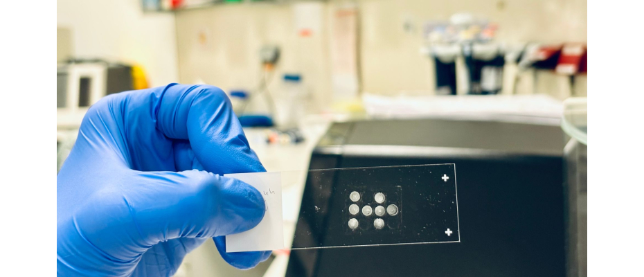
PHOTO
Uniform monolayer of cells anchored onto a glass slide using STAMP, ready for high resolution imaging.