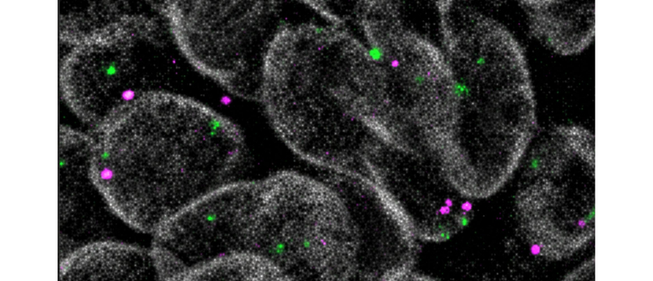
- A new study, published in Science Advances, reveals that the three-dimensional (3D) architecture of the genome actively contributes to tissue regeneration following injury.
- The research has been led by the Faculty of Biology and the Institute of Biomedicine of the University of Barcelona and the Structural Genomics Group at Centre Nacional d’Anàlisi Genòmica (CNAG).
September 17, 2026. When tissues are injured, cells activate complex genetic programmes to repair the damage and restore normal function. Yet how these programmes are orchestrated remains one of the key unanswered questions in biology. Researchers have long known that DNA changes its activity following injury, but it also reshapes the way it folds inside the cell nucleus. As DNA reorganises into a new three-dimensional (3D) arrangement, chromatin loops bring distant parts of the genome together, acting as molecular bridges that allow them to communicate and coordinate tissue repair. A new study, published in Science Advances, has now revealed that the formation of these loops is not merely a consequence of regeneration: it is an essential part of the process.
The research was led by the
Faculty of Biology and the Institute of Biomedicine of the University of Barcelona (IBUB), together with CNAG, in collaboration with the
University of Lausanne (Switzerland).
Using the advanced Hi-C technology available at CNAG, the researchers mapped how the genome reorganises itself in three dimensions during tissue regeneration and identified three chromatin loops that are essential for activating the genetic programme required for tissue repair. When the DNA regions responsible for creating these loops were disrupted, the tissue largely lost its ability to regenerate, while the organism's normal development remained virtually unaffected.
"For decades, we have studied the genome as a linear sequence of DNA. In reality, however, the two metres of DNA inside every human cell are intricately folded into a nucleus only a few micrometres wide. Thanks to technologies that capture the genome's spatial organisation, we can now analyse it much more closely to how it actually exists inside living cells. This study shows that this spatial organisation is not just a way of packaging DNA, but a fundamental mechanism that helps control tissue regeneration", explains Dr Marc Martí-Renom, ICREA Research Professor at CNAG, and the Centre for Genomic Regulation (CRG). The findings identify the 3D architecture of the genome as a previously unrecognised layer of regulation that is key for successful tissue regeneration.
Uncovering the hidden role of genome architecture
Although the importance of genome architecture is increasingly recognised, little is known about how changes in its 3D organisation influence gene activity during tissue regeneration. However, growing evidence suggests that the regenerative capacity of damaged tissues is closely linked to the changes in chromatin. To address this, researchers studied the wing imaginal discs of Drosophila melanogaster (the fruit fly) as a model of tissue regeneration.
"Chromatin adopts a complex three-dimensional architecture that enables interactions between regions of the genome that are located far apart and regulates which genes are activated or remain silent at any given time," explains Professor Montserrat Corominas, from the University of Barcelona's Department of Genetics, Microbiology and Statistics, and corresponding author of the study.
Taken together, the findings provide the first direct evidence that changes in the three-dimensional architecture of the genome actively contribute to tissue regeneration, revealing a previously unknown layer of gene regulation during this process.
REFERENCE ARTICLE
Palmira Llorens-Giralt et al. ,3D genome organization in tissue regeneration involves long-range chromatin loops.Sci. Adv.12,eaea8281(2026).DOI:
10.1126/sciadv.aea8281