- Researchers have discovered a window of opportunity to eliminate metastasis at an early stage, before it causes symptoms.
- The finding, published in Cancer Cell, opens a potential route to stopping the disease before it becomes clinically apparent, effectively paving the way towards prevention. A similar strategy could also be used to prevent relapse after surgery.
- The study was led by the
Spanish National Cancer Research Centre (CNIO), with contributions from CNAG’s Spatial Genomics Team, which provided key spatially-resolved single-cell analyses of patient brain samples with micrometastases.
October 13, 2026. Metastasis, the process by which a tumour spreads throughout the body, is associated with the majority of cancer deaths. The Spanish National Cancer Research Centre (CNIO) has investigated the earliest stages in which metastatic cells establish themselves in distant organs, and has discovered a transient phase during which these metastatic seeds are particularly vulnerable, known as ‘proliferate pause’. The study, published in Cancer Cell, was led by Manuel Valiente, head of CNIO’s Brain Metastasis Group, in collaboration with CNAG and around 20 more institutions from Spain, the United Kingdom, Germany, France and the United States.
One of the most important contributions of the study is its potential clinical implications. According to the authors, the findings “open a window of opportunity to attack the disease before symptoms appear,” says Valiente. “This is the beginning of the race to prevent metastasis.” The results also lay the groundwork for potential strategies to prevent metastasis from recurring after surgical removal.
The researchers have also shown, both in mice and in human samples of brain metastases, that these micrometastases can be eliminated using three drugs, two of which are already approved for other indications. At present, however, this approach is not yet possible in clinical practice, as there are no diagnostic tests capable of detecting micrometastases, nor biomarkers that can reliably identify patients at risk. The researchers’ next step will be to launch clinical trials to assess the effectiveness of the drugs tested and identify potential biomarkers.
A ‘proliferative pause’ in which micrometastases are vulnerable
It was already known that cancer cells that colonise another organ (the brain, for example) must adapt to an environment that is very different from that of the tumour in which they originated, such as a tumour in the lung, breast or skin. This new environment is so hostile that many metastatic cells die during the adaptation process.
What this new research has discovered is that tumour cells that do survive enter a period the authors call a proliferative pause. During this stage, metastatic cells seek ways to survive in the new environment and temporarily abandon their aggressive behaviour.
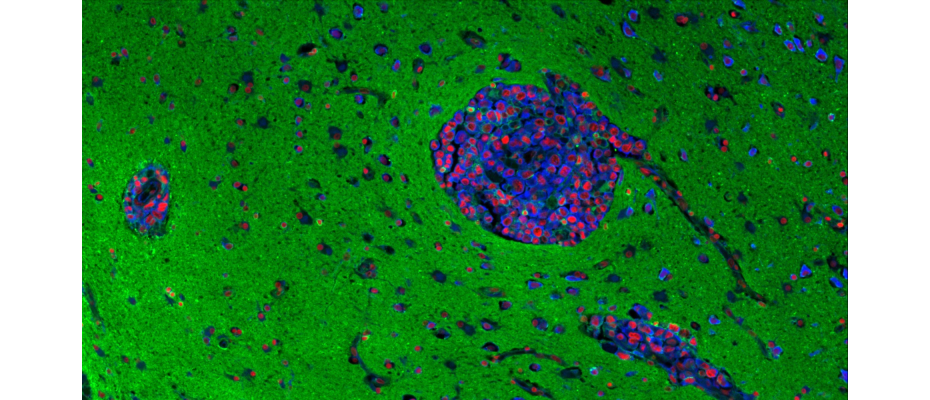
To characterise the behaviour of these metastatic cells in detail, CNAG’s Spatial Genomics Team conducted in situ single-cell analyses of human brain samples containing micrometastases using Xenium (10x Genomics), and interpreted the resulting data. This work helped identify the molecular features of these early metastatic lesions and characterise their state during the proliferative pause.
“Understanding how tumour cells behave in tissues and the mechanisms that regulate them is essential to better understand tumour biology before metastasis develops. Studying these early stages can help us identify the changes that allow cancer cells to adapt and progress, which may help us intervene earlier and ultimately contribute to better outcomes for patients.”, explains Dr Anna Pascual-Reguant, Spatial Genomics Team Leader at CNAG.
The study focused on brain metastasis, although the authors hypothesise, and intend to investigate in future studies, that the vulnerable stage they have identified may be common to the metastatic process more broadly.
REFERENCE ARTICLE
Martínez-Mateu, Sergio H., et al. ‘Uncovering Combination Therapies for Immune-Mediated Inflammatory Diseases through Systems Biology Analysis on Longitudinal Patient Data’. Cell Reports Medicine, Sept. 2026, p. 103026. DOI.org (Crossref),
https://doi.org/10.1016/j.xcrm.2026.103026.
FIGURE
Human brain tissue profiled by Xenium in situ spatial transcriptomics (427-gene panel), in which four neighboring micrometastases are visible as dense nuclear clusters.